Ribosome : Defination, Structure, Functions , Types
Ribosome Definition, structure, functions , types.
Definition of Ribosome
A ribosome is a molecule of macro dimensions that is responsible for the synthesis or translation of the amino acids of mRNA (in eukaryotic cells) and the manufacture of proteins in living beings (in eukaryotic and prokaryotic cells).
What are Ribosomes
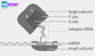
Ribosomes are the most abundant cell organelles and are involved in protein synthesis. They are not surrounded by a membrane and are made up of two types of subunits: a large one and a small one, as a general rule the large subunit is almost twice the small one.
The prokaryotic lineage has 70S ribosomes composed of a large 50S subunit and a small 30S subunit. Likewise, the ribosomes of the eukaryotic lineage are composed of a large 60S subunit and a small 40S subunit.
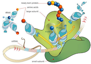
The ribosome is analogous to a factory in motion, capable of reading messenger RNA, translating it into amino acids, and binding them by peptide bonds.
Ribosomes are equivalent to almost 10% of the total protein of a bacterium and more than 80% of the amount of total RNA. In the case of eukaryotes, they are not as abundant compared to other proteins, but their number is greater.
In 1950, researcher George Palade first visualized ribosomes and this discovery was awarded the Nobel Prize in Physiology or Medicine.
Composition of Ribosome
The ribosomes are made up of two subunits, one of proportional size and the other smaller, passing between them is a compressed nucleic acid chain. Each subunit of the ribosome is made up of a ribosomal RNA and a protein. Together they organize the translation and catalyze the reaction to generate polypeptide chains that will be the basis for proteins.
In this same order of ideas, transfer RNAs (tRNAs) are responsible for bringing amino acids to the ribosome and matching the messenger RNA with the amino acids that encode the protein that will be produced by the ribosome.
Characteristics of Ribosome
We distinguish two types of ribosomes
- Ribosomes 70 S and Ribosomes 80 S. Ribosomes 70 S are typical of prokaryotes and of chloroplasts and mitochondria. 80 S ribosomes are typical of eukaryotic cells.
- Ribosomes are made up of two subunits of unequal size and different sedimentation coefficients. One is the largest subunit and the other is the minor subunit.
- The 70 S ribosomes have a larger subunit with a sedimentation coefficient of 50 S and a smaller one of 30 S.
- The 80 S ribosomes have the largest subunit with a 60 S coefficient and the other 40 S.
- The union of both subunits is performed in the presence of a 0.001 M concentration of Mg ions, if this concentration decreases the separation of the two subunits would occur, therefore it is a reversible process.
- If the molar concentration of Mg ions increases up to 10 times, the union of two ribosomes occurs to give rise to the dimers.
- The dimer of 70 S ribosomes has a coefficient of 100 S, whereas, in the dimer of 80 S ribosomes, the sedimentation coefficient would be 120.
- The ultrastructure of the ribosome is very complex, consisting of ribosomal RNA and proteins. There are different types of ribosomal RNA taking into account the sedimentation coefficient.
- If we perform a phenol extraction of two subunits of an R 70 S, the proteins are separated on the one hand and a ribosomal RNA with a sedimentation coefficient of 23 S on the other, and proteins and 16 S ribosomal RNA appear on the 30 S.
What Are Ribosomes Made Of?
Ribosomes consist of about 60 percent protein and about 40 percent ribosomal RNA. This is an interesting relationship since one type of RNA (messenger RNA or RNA) is required for protein synthesis or translation. So, in a way, ribosomes are like a dessert consisting of unmodified cocoa beans and refined chocolate.
RNA is one of two types of nucleic acids found in the world of living things, the other is deoxyribonucleic acid or DNA. DNA is the more notorious of the two, and they are often mentioned not only in major scientific articles but also in crime stories. But RNA is actually the most versatile molecule
Structure of Ribosome
- Ribosomes are small cellular structures (from 29 to 32 nm, depending on the group of the organism), rounded and dense, made up of ribosomal RNA and protein molecules, which they find associated with each other.
- The most studied ribosomes are those of eubacteria, archaea, and eukaryotes. In the first lineage, the ribosomes are simpler and smaller. Eukaryotic ribosomes, meanwhile, are more complex and larger. In archaea, ribosomes are more similar to both groups in certain respects.
- Vertebrate and angiosperm ribosomes (flowering plants) are particularly complex.
- Each ribosomal subunit is primarily made up of ribosomal RNA and a wide variety of proteins. The large subunit may be made up of small RNA molecules, in addition to ribosomal RNA.
- Proteins are coupled to ribosomal RNA in specific regions, in order. Within the ribosomes, several active sites can be differentiated, such as catalytic zones.
- Ribosomal RNA is of crucial importance to the cell and this can be seen in its sequence, which has been virtually unchanged throughout evolution, reflecting the high selective pressures against any change.
Types of ribosomes
Ribosomes in prokaryotes
Bacteria, like E. coli, possess more than 15,000 ribosomes (in proportions this equals almost a quarter of the dry weight of the bacterial cell).
The ribosomes in bacteria are about 18nm in diameter and consist of 65% ribosomal RNA and only 35% proteins of various sizes, between 6,000 and 75,000 kDa.
The large subunit is called the 50S and the small 30S, which combine to form a 70S structure with a molecular mass of 2.5 × 10⁶ kDa.
The 30S subunit is elongated in shape and not symmetrical, while the 50S is thicker and shorter.
The small E. coli subunit is made up of 16S ribosomal RNAs (1542 bases) and 21 proteins and in the large subunit are 23S ribosomal RNAs (2904 bases), 5S (1542 bases), and 31 proteins. The proteins that compose them are basic and the number varies according to the structure.
Ribosomal RNA molecules, along with proteins, are grouped into a secondary structure similar to other types of RNA.
Ribosomes in eukaryotes
Ribosomes in eukaryotes (the 80S) are larger, with higher RNA and protein content. RNAs are longer and are called 18S and 28S. As in prokaryotes, the composition of ribosomes is dominated by ribosomal RNA.
In these organisms, the ribosome has a molecular mass of 4.2 × 10⁶ kDa and breaks down into the 40S and 60S subunits.
The 40S subunit contains a single RNA molecule, 18S (1874 bases), and some 33 proteins. Likewise, the 60S subunit contains the 28S (4,718 bases), 5.8S (160 bases), and 5S (120 bases) RNAs. Furthermore, it is made up of basic proteins and acidic proteins.
Ribosomes in archaea
Archaea are a group of microscopic organisms that resemble bacteria but differ in so many characteristics that they constitute a separate domain. They live in diverse environments and are capable of colonizing extreme environments.
The types of ribosomes found in archaea are similar to the ribosomes of eukaryotic organisms, although they also have certain characteristics of bacterial ribosomes.
It has three types of ribosomal RNA molecules: 16S, 23S, and 5S, coupled to 50 or 70 proteins, depending on the study species. Regarding the size, the ribosomes of archaea are closer to the bacterial ones (the 70S with two subunits 30S and 50S) but in terms of their primary structure, they are closer to the eukaryotes.
Since archaea usually inhabit environments with high temperatures and high salt concentrations, their ribosomes are highly resistant.
Synthesis of Ribosome
All of the cellular machinery necessary for ribosome synthesis is located in the nucleolus, a dense region of the nucleus that is not surrounded by membrane structures.
The nucleolus is a variable structure depending on the cell type: it is large and conspicuous in cells with high protein requirements and it is an almost imperceptible area in cells that synthesize little amount of protein.
Processing of ribosomal RNA occurs in this area, where it couples to ribosomal proteins and gives rise to products of granular condensation, which are the immature subunits that form functional ribosomes.
The subunits are transported outside the nucleus – through the nuclear pores – to the cytoplasm, where they are assembled into mature ribosomes that can start protein synthesis.
What is the Function of Ribosomes?
The main functions of ribosomes can be summarized below:
- They connect amino acids to form specific proteins, proteins are essential to carry out cellular activities.
- In the process of making proteins, deoxyribonucleic acid produces mRNA by the DNA translation process.
- The genetic message of the mRNA is translated into proteins during DNA transcription.
- Protein assembly chains during protein synthesis are defined in mRNA.
- The mRNA is synthesized in the nucleus and transferred to the cytoplasm for an additional process of protein synthesis.
- In the cytoplasm, the two ribosome subunits are coupled around the mRNA polymers; proteins are then synthesized with the help of transfer RNA.
- Proteins that are synthesized by ribosomes present in the cytoplasm are used in the cytoplasm itself. Proteins originating from bound ribosomes are transported outside the cell.
Important points about Ribosomes:
- Almost all the proteins required by cells are synthesized by ribosomes. Ribosomes are ‘free’ in the cell cytoplasm and also bind to the rough endoplasmic reticulum.
- Ribosomes receive information from the cell nucleus and from the building materials of the cytoplasm.
- Ribosomes translate the information encoded into messenger ribonucleic acid (mRNA).
- They bind specific amino acids together to form polypeptides and export these to the cytoplasm.
- A mammalian cell can contain up to 10 million ribosomes, but each ribosome only has a temporary existence.
- Ribosomes can bind amino acids at a rate of 200 per minute.
- Ribosomes are formed by blocking a small subunit into a large subunit. Subunits are normally available in the cytoplasm, with the largest being about twice the size of the smallest.
- Each ribosome is a complex of ribonucleoproteins with two-thirds of its mass is composed of ribosomal RNA and about a third of ribosomal protein.
- Protein production takes place in three stages: initiation, (2) elongation, and (3) termination.
- During the production of peptides, the ribosome moves along the mRNA in an intermittent process called translocation.
- Antibiotic drugs, such as streptomycin, can be used to attack the translation mechanism in prokaryotes. This is very useful. Unfortunately, some bacterial toxins and viruses can also do this.
- After leaving the ribosome, most proteins fold or are modified in some way. This is called “post-translational modification.”
Frequently Asked Questions
- A look at the protein production line that can bind amino acids at a rate of 200 per minute!
- In the ribosome, there are THREE STAGES and THREE operating SITES involved in the protein production line.
- The three STAGES are (1) Initiation, (2) Elongation, and (3) Termination.
- The three operational or binding SITES are A, P, and E that are read from the mRNA entry site (conventionally the right side).
- Sites A and P encompass both ribosome subunits with a larger portion residing in the large ribosome subunit and a smaller portion in the smaller subunit. Site E, the exit site, resides in the large ribosomal subunit.


