Maharashtra Board Class 12 Biology Chapter 2- Reproduction in lower & higher Animals Solutions
Q. 1 Multiple Choice Questions
1. The number of nuclei present in a zygote
is ……
a. two
b.one
c. four
d.eight
2. Which of these is the male reproductive organ in human?
a. sperm
b. seminal fluid
c. testes
d. ovary
3. Attachment of embryo to the wall of the uterus is known as……..
a. fertilization
b. gestation
c. cleavage
d. implantation
4. Rupturing of follicles and discharge of ova is known as …………..
a. capacitation
b. gestation
c. ovulation
d. copulation
5. In human female, the fertilized egg gets implanted in uterus ………………
a. After about7 days of fertilization
b. After about 30 days of fertilization
c. After about two months of fertilization
d. After about 3 weeks of fertilization
6. Test tube baby technique is called………
a. In vivo fertilization
b. In situ fertilization
c. In vitro fertilization
d. Artificial insemination
7. The given figure shows a human sperm. Various parts of it are labelled as A, B, C, and D. Which labelled part represents acrosome?
a. B
b. C
c. D
d. A
8. Presence of beard in boys is a …………
a. primary sex organ
b. secondary sexual character
c. secondary sex organ
d. primary sexual character
Q.2: Answer in One Sentence
Q. 4 Short Answer Questions.
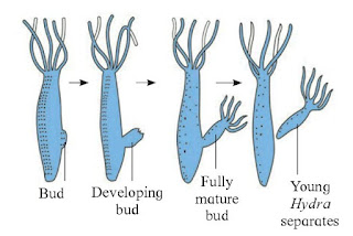 |
| Fig: Budding in Hydra |
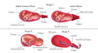 |
| Fig : Parturition |
2. Expulsion stage : The uterine and abdominal contractions become stronger. In normal delivery, the foetus passes out through cervix and vagina with head in forward direction. It takes 20 to 60 min. The umbilical cord is tied and cut off close to the baby’s navel.
3. After birth : After the delivery of the baby the placenta separates from the uterus and is expelled out as “after birth”, due to severe contractions of the uterus. This process happens within 10 to 45 minutes of delivery.
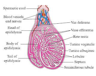 |
| Fig : Testis Structure. |
The testis is externally covered by a collagenous connective tissue layer called tunica albuginea. Outer to it is an incomplete peritoneal covering called tunica vaginalis, and inner to it is tunica vasculosa, a thin membranous and vascular layer. Fibers from tunica albuginea divide each testis into about 200-300 testicular lobules. Each with 1-4 highly coiled seminiferous tubules. Each seminiferous tubule is internally lined by cuboidal germinal epithelial cells (spermatogonia) and few large pyramidal cells called Sertoli or sustentacular cells.
The germinal epithelial cells undergo gametogenesis to form the spermatozoa. Sertoli cells provide nutrition to the developing sperms. Various stages of spermatogenesis can be seen in the seminiferous tubules. The inner most spermatogonial cell (2n), primary spermatocyte (2n), secondary spermatocyte (n), spermatids (n) and sperms (n). The Interstitial or Leydig’s cells lie in between the seminiferous tubules. They secrete the male hormone androgen or testosterone.
4. Describe the structure of blastula.
Answer :
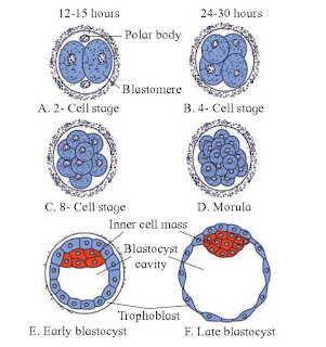 |
| Fig: Cleavage & Blastula Formation |
The development of multi-cellular organisms started from fertilization. Fertilization forms a single-celled zygote, which undergoes rapid cell division to form the blastula. The rapid, multiple rounds of cell division are termed cleavage. The cleavage has produced over 80-100 cells, the embryo is called a blastula. The blastula is usually a spherical layer of cells (the blastoderm) surrounding a fluid-filled or yolk-filled cavity (the blastocoel). Mammals at this stage form a structure called the blastocyst, characterized by an inner cell mass that is distinct from the surrounding blastula,
During cleavage, the cells divide without an increase in mass; that is, one large single-celled zygote divides into multiple smaller cells. Each cell within the blastula is called a blastomere.
In mammals, the blastula forms the blastocyst in the next stage of development. Here the cells in the blastula arrange themselves in two layers: the inner cell mass, and an outer layer called the trophoblast. The inner cell mass is also known as the embryoblast and this mass of cells will go on to form the embryo. At this stage of development, the inner cell mass consists of embryonic stem cells that will differentiate into the different cell types needed by the organism. The trophoblast will contribute to the placenta and nourish the embryo.

The ovary is an organ found in the female reproductive system that produces an ovum. It is the primary female sex organ. Its main function is production of egg or ovum and the female reproductive hormones. It is solid, oval or almond shaped organ. It is 3.0 cm in length, 1.5 cm in breadth and 1.0 cm thick. It is located in the upper lateral part of the pelvis near the kidneys. Each ovary is a compact structure differentiated into a central part called medulla and the outer part called cortex.
6. Describe the various methods of birth control to avoid pregnancy.
Answer : The birth control measures which prevent fertilization are referred to as contraceptives. The contraceptive methods help to prevent unwanted pregnancies. An ideal contraceptive should be easily available, user friendly, effective and with no or least side effects.
A) Temporary and
B) Permanent.
Temporary methods:
These are of following types :
1. Natural method/ Safe period / Rhythm method .
2. Coitus Interruptus or withdrawal.
3. Lactational amenorrhea (absence of menstruation).
4. Chemical means (spermicides).
5. Mechanical means / Barrier methods: In this method, with the help of barriers the ovum and sperm are prevented from physically meeting. These mechanical barriers are of three types. i) Condom, ii) Diaphragm, cervical caps and vaults, iii) Intra-uterine devices (IUDs)
6. Physiological (Oral) Devices : Physiological devices are used in the form of tablets and hence are popularly called pills. It is an oral contraceptive, used by the female.
B. Permanent Method:
The permanent birth control method in men is called vasectomy and in women it is called tubectomy.
These are surgical methods, also called sterilization. In vasectomy a small part of the vas deferens is tied and cut where as in tubectomy, a small part of the fallopian tube is tied and cut. This blocks, gamete transport and prevent pregnancy.
1. Medical Termination of Pregnancy (MTP) : An intentional or voluntary termination of pregnancy before full term is called Medical termination of Pregnancy (MTP) or induced abortion. MTP is essential in cases of unwanted pregnancies or in defective development of foetus. It is safe during the first trimester of pregnancy. The defective development of foetus is examined by amniocentesis.
Answer : Goals of RCH Programmes:
2. To provide the facilities to people to understand and build up reproductive health.
3. To provide support for building up a reproductively healthy society.
4. To bring about a change mainly in three critical health indicators i.e. reducing total infertility rate, infant mortality rate and maternal mortality rate.
Answer : Parturition is the process of giving birth to a baby as the development of the foetus gets completed in the mother’s womb. The hormones involved in this process are oxytocin and relaxin. Oxytocin leads to the contraction of smooth muscles of myometrium of the uterus, which directs the full term foetus towards the birth canal. On the other hand, relaxin hormone causes relaxation of the pelvic ligaments and prepares the uterus for child birth
Answer : The male accessory ducts (vasa efferentia, epididymis, vas deferens, and rete testis) serve to temporarily store spermatozoa and to transport them outside urethra during ejaculation.
Answer : Sperm capacitation refers to the physiological changes spermatozoa must undergo in order to have the ability to penetrate and fertilize an egg. Recognition of the phenomenon was quite important to early in vitro fertilization experiments as well as to the fields of embryology and reproductive biology.
Q. 6 Long answer questions.
Answer : Male Reproductive System :
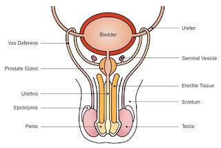 |
| Fig : Male reproductive system |
It consists of the primary male organ (gonad) called testes, the accessory ducts and glands which form internal and external genitalia.
Testis : A pair of testes, mesodermal in origin, are formed in lower abdominal cavity. They are located in a pouch called scrotum. During early foetal life, the testes develop in abdominal cavity and later they descend into the scrotal sac through a passage called inguinal canal. Each testis is oval in shape, 4 to 5cm long, 2 to 3cm wide and 3cm thick.
Seminal vesicles: It is a pair of glands lying on the posterior side of urinary bladder. It secretes an alkaline seminal fluid which contains fructose, fibrinogen and prostaglandins. It contributes about 60% of the total volume of the semen. Fructose provides energy for sperm movement while fibrinogen coagulates the semen into a bolus for quick propulsion in the vagina.
Prostate gland: It is a large and single gland made up of 20-30 lobes and is located underneath the urinary bladder. It surrounds the urethra and releases a milky white and slightly acidic prostatic fluid into the urethra. It forms about 30% of volume of semen. It contains citric acid, acid phosphatase and various other enzymes. The acid phosphatase protects the sperms from the acidic environment of vagina.
The accessory ducts include rete testis, vasa efferentia, epididymis, vas deferens, ejaculatory duct and urethra. All the seminiferous tubules of the testis at the posterior surface form a network of tubules called rete testis. 12-20 fine tubules arising from rete testis are vasa efferentia. They carry the sperms from the testis and open into the epididymis.
Answer : Female Reproductive System: The female reproductive system consist of the following parts :
- a. Infundibulum : The proximal funnel like part with an opening called ostium surrounded by many finger like processes called fimbriae (of these at least one is long and connected to the ovary). The cilia and the movement of fimbrae help in driving the ovulated egg to the ostium.
- b. Ampulla : It is the middle, long and straight part of the oviduct. Fertilization of the ovum takes place in this region.
- c. Isthmus / Cornua : The distal narrow part of the duct opening into the uterus.
3. Uterus : It is commonly also called the womb. It is a hollow, muscular, pear shaped organ, located above and behind the urinary bladder. It is about 7.5 cm long, 5 cm broad and 2.5 cm thick. The uterus can be divided into three regions :
- a. Fundus : It is the upper dome shaped part. Normally implantation of the embryo occurs in the fundus.
- b. Body : It is the broad part of the uterus which gradually tapers downwards.
- c. Cervix : It is the narrow nec about 2.5 cm in length. It extends into the vagina. Its passage has two openings : an internal os towards the body, and an external os towards the vagina.
4. Vagina : It is a tubular, female copulatory organ, 7 to 9 cm in length. It lies between the cervix and the vestibule. The vaginal wall has an inner mucosal lining, the middle muscular layer and anouter adventitia layer. The mucosal epithelium is stratified and non-keratinised and stores glycogen. The opening of the vagina into the vestibule is called vaginal orifice. This opening is covered partially by a fold of mucus membrane called hymen. The vagina acts as a passage for menstrual flow as well as birth canal during parturition.
5. External genitalia : The external genital organs of female include parts external to the vagina and are collectively called ‘vulva’ (covering or wrapping), or pudendum.
6. Accessary glands / Vestibular glands / Bartholin’s glands : It is a pair of glands homologous to the Bulbourethral or Cowper’s glands of the male. They open into the vestibule and release a lubricating fluid.
Answer : Fertilization / Syngamy:
Sexual reproduction primarily involves formation and fusion of gametes. Fertilization is the later process which involves fusion of the haploid male and female gametes resulting in the formation of a diploid zygote (2n). Like in other mammals, in humans the process of fertilization is internal and it usually takes place in the ampulla of the fallopian / uterine tube. The fertilized egg or zygote will develop into an embryo and this process occurs within the uterus.
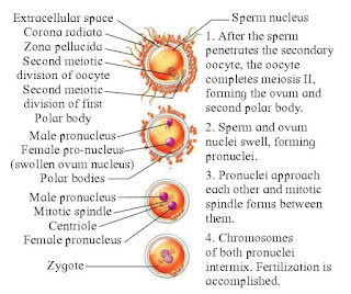 |
| Fig : Process of Fertilization |
Mechanism of fertilization : Semen released during ejaculation has sperms and some secretions. The coagulated semen now undergoes liquification and sperms become active. The mechanism of fertilization is as follows :
Movement of sperm towards egg: It involves capacitation of sperms reaching the vagina. Here as many as 50% are demotilised / broken / destroyed. Remaining sperms undergo capacitation. This process requires 5-6 hours.
Due to the acrosomal reactions, the plasma membrane of the secondary oocyte and the sperm fuse together so that the contents of the sperms can enter. When the plasma membrane of the sperm binds with that of the secondary oocyte, the plasma membrane of the oocyte depolarizes. This prevents polyspermy.
Calcium ions play a significant role in the acrosomal reaction. The main factors essential for acrosomal reactions are optimum pH, temperature and calcium and magnesium concentration.
Cortical Reaction : Soon after the fusion of the plasma membranes, the oocyte shows cortical reactions. Cortical granules found under the plasma membrane of the oocyte, which fuses with the plasma membrane and releases cortical enzymes between the zona pellucida and plasma membrane. The zona pellucida is hardened by the cortical enzymes that prevent polyspermy.
Sperm Entry into the ovum /egg : A projection known as the cone of reception is formed by the secondary oocyte at the point of sperm contact. This cone of reception receives the sperm.
Karyogamy : After the entry of the sperm, the suspended second meiotic division is completed by the secondary oocyte. This gives rise to a haploid ovum and a second polar body. The head of the sperm containing the nucleus detaches from the entire sperm and is known as male pronucleus. The tail and the second polar body degenerates. The nucleus of the ovum is known as female pronuclei.
The male and female pronuclei fuse and their nuclear membranes degenerate. The fusion of the chromosomes of male and female gametes is called karyogamy. The ovum is now fertilized and is known as a zygote.
Activation of Ovum / Eggs: The entry of sperm triggers the metabolism in the zygote. Consequently, protein synthesis and cellular respiration increase.
Fusion of egg and sperm : The coverings of male and female pronuclei degenerate allowing the chromosomal pairing. This results in the formation of a synkaryon by theprocess called syngamy or karyogamy. The zygote is thus formed.
Implantation : Once fertilization happens, the cell starts to divide and multiply within 24 hours in the fallopian tube. This detached multi-celled structure is called zygote. Later, after 3-4 days it travels to the uterus and now we call it as an embryo. The embryo develops and undergoes various stages and gets attached to the endometrial layer of the uterus. This process of attachment is known as implantation.
| Chapter 2: Reproduction in Lower & Higher Animals |
| Chapter 3: Inheritance and Variation |
| Chapter 4: Molecular Basis of Inheritance |
| Chapter 6: Plant Water Relation |
| Chapter 7: Plant Growth and Mineral Nutrition |
| Chapter 8: Respiration and Circulation |
| Chapter 9: Control and Coordination |
| Chapter 10: Human Health and Diseases |
| Chapter 11: Enhancement of Food Production |
| Chapter 12: Biotechnology |
| Chapter 13: Organisms and Populations |
| Chapter 14: Ecosystems and Energy Flow |

